

关键字: 3D Microendoscopy 发布时间:2011-4-12
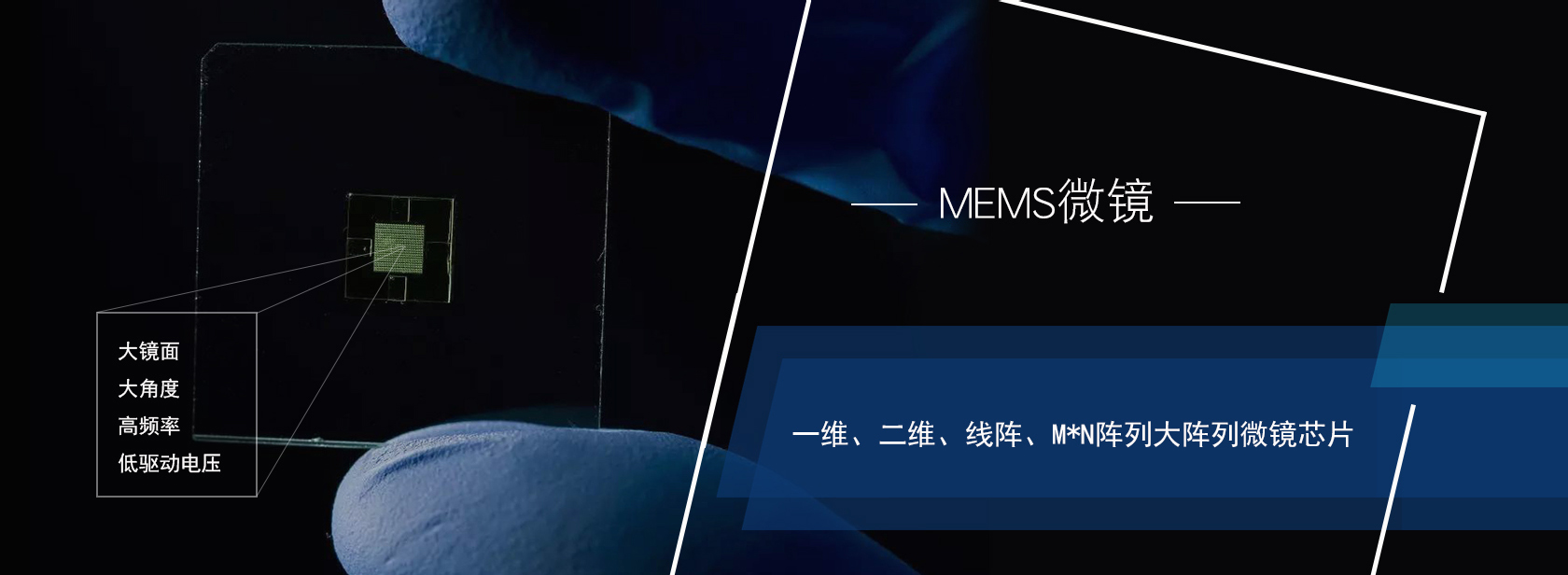
我公司领军人应苏州大学邀请,于4月13日下午在苏州大学电子信息楼为苏大学子做一份关于“微型内窥镜三维实时成像技术”的报告,届时谢博士将介绍MEMS加工技术、MEMS微镜和微透镜等微型光学器件。这些微型光学器件使无损实时“光学切片”内窥镜三维影像成为可能,这是内窥镜技术的一次巨大变革。另外,还将特别介绍基于MEMS的光相干断层扫描(OCT),共聚焦和非线性光学内窥镜影像技术及其在早期癌症诊断和肿瘤精准切除手术方面的应用。 Over 7 million people die of cancer worldwide each year. The high mortality is mainly due to the lack of early cancer detection modalities especially for internal organs. CT, MRI and ultrasound imaging have issues of low resolution, low contrast, safety, or high cost. Several optical imaging techniques provide high-resolution cross-sectional information suitable for in vivo noninvasive early cancer diagnosis. However, these optical imaging systems are typically bulky and slow, and thus are difficult to apply to internal organs where most cancers are originated. Microelectromechanical systems (MEMS) technology offers the advantages of small size and fast scan speed, providing a tremendous opportunity for realizing real-time in vivo endoscopic imaging. In this talk, a unique MEMS technology that can create large-range, multi-axis, rapid scanning micromirrors and microlenses will be introduced. These MEMS devices in turn enable endoscopic optical “biopsy” modalities, resulting in a paradigm shift of optical imaging of internal organs. In particular, MEMS based 3D endoscopic optical coherence tomography (OCT) imaging, nonlinear optical imaging and confocal imaging will be introduced and in vivo experimental results of animals will be presented. |
2018无锡微文半导体科技有限公司 版权所有 苏ICP备16003840号-2

中国・ 无锡新区菱湖大道200号
中国无锡传感国际创新园B2-305
电话:13358103275